한국수의안과인증의 시험 규정집 (Bylaws of Korean Society of Veterinary Ophthalmology)
규정선정 위원서강문, 유석종, 지동범, 정만복, 김준영


- 가.한국수의안과연구회는 수의안과인증의 시험을 매년 시행하며, 시험 지원자의 최소 자격요건은 아래와 같이 정한다.
- 나.한국수의안과인증의 시험은 필기시험(Written examination and Slide Recognition test)과 실기시험(Practical and Oral test)으로 나누어 실시하며, 상세한 규정은 아래와 같이 정한다.
- 다.실기시험은 필기시험을 통과한 응시자에 한하여 실시한다.
Part I시험 지원요건
- 가.한국에서 수의사면허를 취득하였으며 한국 수의안과연구회 정회원인 수의사
- 나.안과에 관한 연구, 출판 및 강의를 통하여 한국 수의안과 발전에 실질적으로 기여한 수의사
- 다.수의안과진료 경험이 최소 5년 이상이며 본인 전체 진료시간의 60%를 안과에 분담해야 하며, 이 중에서 50%는 안과 진료에, 나머지 10%는 연구와 강의에 분담해야 한다.
- 라.최근 3년 동안 매년 최소 초진 200 개의 안과진료 경험이 있는 수의사
- 마.한국 수의안과연구회가 정한 최소한의 안과 진단 장비를 갖춘 병원에서 수의안과 진료 경험이 최소 3년 이상인 수의사
- 바.최소 3개 이상의 peer-reviewed 안과관련 논문을 제 1저자 및 교신저자로 출판한 (이 중에서 최소 한편은 SCI 혹은 SCI-E급에 출판) 수의사
(종설과 초록은 포함하지 않는다) - 사.최소 3개 이상의 안과연구를 1저자 및 교신저자로 안과관련학회에 발표한 수의사. 이중에서 반드시 한편은 국제수의안과관련학회 (ACVO, ECVO, AiCVO)에 발표해야 한다.
Part II시험범위 및 추천서적목록
- 가.시험범위 : 한국수의안과인증의 시험 범위는 아시아수의안과전문의 시험 범위를 기준으로 하며, 아래의 list를 최소규정으로 하여 실시한다.
1. List of Required Textbooks.
-
(1)Gelatt KN., et al. Veterinary Ophthalmology (6th edition). Wiley-Blackwell 2021. (Chapter 1 by Cook CS., and chapter 2 by Sauelson DA.)
--Volume 1: The Whole
--Volume 2: Chapter 21-28, Chapter 36, Chapter37 (part 1, part 2) - (2) Gilger BC. Equine Ophthalmology 4th edition. Wiley-Blackwell, 2022.
- (3) Dubielzig R, et al. Veterinary Ocular Pathology: A Comparative Review. Elsevier, 2010.
- (4) Eisner, Georg, Schneider, Peter, Telger, Terry C. Eye Surgery : An Introduction to Operative Technique. Springer, 1990
2. List of Textbooks Recommended for Image Recognition exam preparation
The following texts may be useful in preparing for the Image Recognition portion of the examination. No questions will be derived from these texts for the Written portion of the examination.
- (1) Barnett KC. Color Atlas of Veterinary Ophthalmology, Williams and Wilkins, (Most recent edition).
- (2) Barnett KC. Color Atlas and Text of Equine Ophthalmology, CV Mosby/Wolf, (Most recent edition).
- (3) Ketring KL., and Glaze MB. Atlas of Feline Ophthalmology. Veterinary Learning Systems, (Most recent edition).
- (4) Dziezyc J., and Nicholas JM. Color Atlas of Canine and Feline Ophthalmology. W.B. Saunders (Most recent edition).
- (5) Lim CC. Small Animal Ophthalmic Atlas and Guide. Wiley-Blackwell. (Most recent edition).
3. List of Required Journals Past seven years of journal articles in print prior to January 1 of the year of the examination. Date of e-publication is irrelevant. Example: The hard copy publication dates covered on the exam range from January 1, 2009-December 31, 2015. (Note: Beginning with journal publication dates of January 1, 2010 and later, no questions on the Written examination will be derived from case reports that involve single animals. Review of images in these case reports is recommended for Image Recognition exam preparation.)
3.1 Essential Veterinary Journals
The large majority of questions for the written examination will be derived from these journals.
- (1) Veterinary Ophthalmology
- (2) American Journal of Veterinary Research*
- (3) Equine Veterinary Journal*
- (4) Journal of Small Animal Practice*
- (5) Journal of the American Animal Hospital Association*
- (6) Journal of the American Veterinary Medical Association*
- (7) Veterinary Clinics of North America*
- (8) Veterinary Pathology*
- (9) Veterinary Record*
- (10) Veterinary Surgery*
- (11) Journal of Veterinary Internal Medicine*
- (12) Veterinary Immunology and Immunopathology* (Note*: Articles from these veterinary journals should be reviewed for any situation or disease that involves ocular, periocular, or neuro-ophthalmic structures or systemic conditions relevant to ophthalmic disease.)
3.2 Human Journals
Questions derived from pertinent articles* from these journals may occur infrequently on the Written examination):
- (1) Current Eye Research
- (2) Experimental Eye Research
- (3) Graefe’s Archives for Clinical and Experimental Ophthalmology
- (4) Investigative Ophthalmology and Visual Science
- (5) Journal of Ocular Pharmacology and Therapeutics (*Note: Review of basic science, human clinical and other veterinary specialty journals should be limited to those articles dealing with situations or diseases directly applicable to veterinary ophthalmology (eg. where a common domestic animal is used as an animal model). Review of human clinical conditions or basic science articles unrelated to veterinary ophthalmology is not necessary for exam preparation.)
3.3 Classical papers
- (1) Veterinary Clinics of North America (All issues and sections specifically relating to ophthalmology from the year 2000 to present. Individual issues/sections specified elsewhere in this reading list are also recommended.)
- (2) Veterinary Clinics of North America 27:5. Surgery Management of Ocular Disease. September, 1997.
Part III시험 형식 및 유형
시험은 총 3개의 part로 나누어져 있고, written examination, Slide Recognition test, Practical and Oral test 로 구성된다. Written examination 100점, slide recognition test 100점, Practical and Oral test 100점으로 구성되며, 각각의 영역에서 70점이상을 넘어야 통과하는 것으로 한다. Practical and Oral test는 3개의 section으로 구성되고 3개의 section의 평균을 이 영역의 점수로 한다. 각 section은 100점 만점으로 하고 section 당 70점이상이 되어야 통과하는 것으로 한다.
-
가.Written Examination
Written examination은 다지 선다형으로 구성되며 총 100문제로 이루어진다. 시험문제는 수의안과학에서 다루는 기초과학부분과, 수의안과학에서 필요한 anatomy/embryology, physiology, pharmacology, microbiology, histopathology, cytology, toxicology, immunology, molecular biology, genetics, medicine, surgery, diagnostics, diagnostic imaging, neuro-ophthalmology가 포함되며, 시험대상이 되는 동물은 canine, feline, equine, large and small ruminant, poultry, laboratory animal, exotic animal, wildlife species 가 총 망라된다. 하지만 시험대상 동물은 exam specification blueprint에 맞추어서 그 비율을 정한다. 시험시간은 별도로 마련된 exam timetable 에 따라 실시되며, 이중 written examination은 session1 과 session2로 구성되며, 각각의 session마다 50문제씩 2시간에 걸쳐 실시되도록 한다.
모든 문제는 한국수의안과학회에서 정하는 수련의 reading list를 기준으로 하여, 저널, 교과서 그리고 대표적인 논문을 근거로 하여 출제한다.
Sample MCQ
다음은 문제로 나올 수 있는 유형이다. 답은 문제의 끝에 있다.
1. Posterior polar subcapsular cataracts에 대한 설명 중 맞는 것은?
- a. 몇몇 품종에서의 유전적인 질환이며, 가장 빈번이 일어나는 품종은 Labrador 품종이다.
- b. 당뇨성 백내장에서 가장 빈번하게 나타난다.
- c. lens instability가 발생하는 전구증상이다.
- d. Progressive retinal atrophy가 있는 개에서 백내장이 발생하는 형태이다.
2. hypertensive retinal detachment는 주로 어떤 type으로 발생하는가?
- a. rhegmatogenous
- b. bullous
- c. traction
- d. 개의 고혈압환자에서는 망막박리가 잘 발생되지 않는다.
3. Bussieres등의 2004년 연구에 따르면 각막궤양 수술에서 porcine small intestinal submucosa (SIS) 사용하는데 있어 적절한 서술은?
- a. SIS는 거부반응이 심해서 고양이에서 사용하는 것이 적절하지 않다.
- b. 말의 각막궤양에서 SIS의 이식으로 발생할 수 있는 복합증은 Progressive keratomalacia, chronic aqueous humor leakage, endophthalmitis, 시력소실이 있을 수 있다.
- c. SIS의 collagen structures는 collagenases에 매우 약하여서 keratomalacia에는 사용할 수 없다.
-
d. SIS는 개의 각막에서 거부반응이 적고, 세균감염이 잘 없어서 각막궤양 수술시 새로운 이식물질로 매우 효과적이고 안전한 물질이다.
- 1.a
- 2.b
- 3.c
-
나.Slide Recognition test
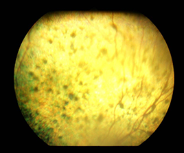
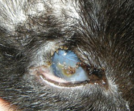
Slide recognition test는 powerpoint-projected images 혹은 color printed image를 통해 문항이 구성되며 총 100문제로 구성된다. 시험은 수의안과학에서 필요한 임상진단부분과 수의안과학과 연관된 모든 images를 포함한다. 따로 현미경을 준비하여 시험을 치르지 않고, 이에 관련된 images 또한 모두 영상으로 만들어 powerpoint-projected images로 시험으로 평가하는 것을 원칙으로 한다. 그러므로 slide recognition test 에는 수의안과학과 연관된 기초과학, diagnostic image, 세포학, 미생물학, 조직병리학을 총망라하여 평가한다. 이 시험은 2개의 sections 으로 하여 이루어지며 각 section마다 50개의 clinical case images로 구성되며 각 image 당 1-4개의 질문으로 구성된다.
문제는 image에 대해 identification, assessment, interpretation을 평가하고 이에 대한 정보를 평가하는 것으로 한다. Slide Recognition test 는 단답형 형태의 답을 원칙으로 하고, 짧은 형태의 서술형 답이 있을 수 있다.
Sample Slide Recognition question
다음은 문제의 유형을 제시한 형태이다.

- 1. 위의 사진에서 병변을 기술하시오
- 2. 위의 안저사진으로 생각할 수 있는 DDx를 기술하시오.

- 1. 위 사진의 병변을 설명하고 진단명을 쓰시오
- 2. 이 질환의 수술방법을 나열하시오.
-
다.Practical and Oral test
Practical and Oral test는 2개로 나누어져 실시된다. 여기엔 환자를 검사하는 techniques과 수술하는 techniques을 평가하며, 이를 수행하는데 필요한 여러가지 지식을 평가하는 것을 목적으로 한다. 2개의 부분은 (1) Extraocular and Corneal surgery (2) Intraocular surgery (ECCE)로 구성된다.
-
(1) Extraocular and Corneal surgery (100 points total)
Extraocular and corneal surgery에서는 cadaver (dog, cat, pig, rabbit등)의 눈을 이용하여 시험이 이루어진다. 수험생은 extraocular나 corneal에 관련된 수술을 하도록 감독관에게 요구받게 되고 이를 수행한다. 필요에 따라 수험생이 자신의 확대할 수 있는 기구를 사용할 수 있다. 수험생은 자신이 수술을 수행하는데 필요한 수술기구와, 여러 소모품을 시험 주체측에 요구할 수 있고 필요에 따라 자신이 사용하는 도구를 감독관 허가하에 사용할 수도 있다. 수술은 50분에 걸쳐 이루어진다.
Adnexal procedures에는 entropion, ectropion 교정술, 안검열상 교정술, eyelid tumor 제거술, 3안검에 관한 수술 그리고 enucleation 중에서 출제가 된다.
Corneal surgery에서는 corneal laceration repair, conjunctival pedicle graft, corneoconjunctival transposition graft, lamellar keratectomy 중에서 출제가 된다. -
(2) Intraocular surgery (100 points total)
Intraocular surgery는 extracapsular extraction (ECCE)를 50분동안 실시하여 평가한다. 모든 수술은 cadaver인 pig eye을 통해서 실시되는 것을 원칙으로 한다. 수술에 필요한 수술용 현미경은 시험주최측에서 제공하여야 한다. 수험생은 수술에 필요한 수술기구와, 여러 소모품을 시험 주최측에 요구할 수 있고, 필요에 따라 자신이 사용하는 도구를 감독관 허가하에 사용할 수도 있다. 수험생은 최근에 사용되고 있는 수술 techniques을 사용하여야 하고 수험생이 가장 많이 익힌 수술 방법에 따라 실시하면 된다. 수험생은 two-step clear corneal incision, continuous curvilinear capsular capsulorhexis, extracapsular extraction, demonstrate cortical removal, closure of the corneal incision을 수행하여야 한다. 감독관은 수험생에게 각각의 수술절차들에 대해 질문하고 그 방법들에 대해 정확히 이해하고 있는지 평가한다. 감독관 중 한명은 수술이 진행되는 동안 surgical assistant로 수술을 도울 수 있으나 수험생이 요구하는 부분만 수행하여야 한다. Cadaver eye는 수술 하루 전 혹은 당일에 도살된 돼지의 눈을 사용하며, 가능하면 당일 아침에 도살된 돼지의 눈을 사용하는 것을 원칙으로 한다. 수술을 시작하면서 돼지의 눈이 tension을 잃어 너무 soft한 경우 이를 보정하기 위해 normal saline을 눈 안에 주사기로 주입하여 일정한 tension을 가할 수 있다. 수술 technique에 대한 기준은 Eisner의 Eye surgery, An introduction to Operative Technique (Springer-Verlag)에 ECCE procedure를 기준으로 한다.
모든 수험생들은 반드시 다양한 종류의 미세수술기구에 대해 충분히 인지하고 있어야 하며, 안내 수술에 대한 기본개념을 숙지하고 있어야 한다. 예를 들면 corneal incision을 봉합하는 방법으로 다양한 수술 pattern이 있고 이들에 대한 적절한 지식을 가지고 있어야 한다. 수험생들은 먼저 적절한 수술방법에 대해 평가 받게 되고 다음 적절한 기구사용에 대해 평가 받게 될 것이다.
모든 2개의 section은 모두 통과하여야 하며, 통과를 하지 못하였다면, 통과하지 못한 section만 다음시험에서 다시 치를 수 있다.
Part IV시험 specification blueprint
| I. Basic Ophthalmology (10% of Exam) | Total No.: 10 | By Region | |
|---|---|---|---|
| 1A Vision Sciences (40%) | 4 | 4 | |
| 1.a Embryology, Anatomy | 2 | ||
| 2.b Physiology and Optics, | 2 | ||
| 1B Clinical ophthalmology (60%) | 6 | 6 | |
| 1.a Immunity | 1 | ||
| 2.b Microbiology | 1 | ||
| 3.c Pharmacology | 1 | ||
| 4.d Pathology | 1 | ||
| 5.e Diagnostic Examination | 2 | ||
| Total No. of Exam Questions | 10 | 10 | |
| II. Canine Ophthalmology(55% of Exam) | Total No.: 55 | By Region | |
|---|---|---|---|
| 2A Adnexa (17%) | 9 | 9 | |
| 1.a Examination | 3 | ||
| 2.b. Assessment | 3 | ||
| 3.c. Treatment planning | 3 | ||
| 2B Anterior Segment (68%) | 38 | 38 | |
| 2B1 cornea (24%) | 13 | ||
| 1.a Examination | 4 | ||
| 2.b. Assessment | 4 | ||
| 3.c. Treatment planning | 5 | ||
| 2B2 iris/CB (10%) | 6 | ||
| 1.a Examination | 2 | ||
| 2.b. Assessment | 2 | ||
| 3.c. Treatment planning | 2 | ||
| 2B3 Glaucoma (15%) | 8 | ||
| 1.a Examination | 2 | ||
| 2.b. Assessment | 3 | ||
| 3.c. Treatment planning | 3 | ||
| 2B4 Lens (19%) | 11 | ||
| 1.a Examination | 4 | ||
| 2.b. Assessment | 3 | ||
| 3.c. Treatment planning | 4 | ||
| 2C Posterior Segment (15%) | 8 | ||
| 1.a Examination | 3 | ||
| 2.b. Assessment | 3 | ||
| 3.c. Treatment planning | 2 | ||
| Total No. of Exam Questions | 55 | 55 | |
| III. Feline Ophthalmology (25% of Exam) | Total No.: 25 | By Region | |
|---|---|---|---|
| 2A Adnexa (17%) | 4 | 4 | |
| 1.a Examination | 1 | ||
| 2.b. Assessment | 1 | ||
| 3.c. Treatment planning | 2 | ||
| 2B Anterior Segment (68%) | 17 | 17 | |
| 2B1 cornea (24%) | 9 | ||
| 1.a Examination | 3 | ||
| 2.b. Assessment | 3 | ||
| 3.c. Treatment planning | 3 | ||
| 2B2 iris/CB (10%) | 4 | ||
| 1.a Examination | 2 | ||
| 2.b. Assessment | 1 | ||
| 3.c. Treatment planning | 1 | ||
| 2B3 Glaucoma (15%) | 2 | ||
| 1.a Examination | 1 | ||
| 2.b. Assessment | 0 | ||
| 3.c. Treatment planning | 1 | ||
| 2B4 Lens (19%) | 2 | ||
| 1.a Examination | 1 | ||
| 2.b. Assessment | 0 | ||
| 3.c. Treatment planning | 1 | ||
| 2C Posterior Segment (15%) | 4 | 4 | |
| 1.a Examination | 2 | ||
| 2.b. Assessment | 1 | ||
| 3.c. Treatment planning | 1 | ||
| Total No. of Exam Questions | 25 | 25 | |
| Iv. Equine Ophthalmology (10% of Exam) | Total No.: 10 | By Region | |
|---|---|---|---|
| 2A Adnexa (17%) | 2 | 2 | |
| 1.a Examination | 1 | ||
| 2.b. Assessment | 0 | ||
| 3.c. Treatment planning | 1 | ||
| 2B Anterior Segment (68%) | 7 | 7 | |
| 2B1 cornea (24%) | 4 | ||
| 1.a Examination | 2 | ||
| 2.b. Assessment | 1 | ||
| 3.c. Treatment planning | 1 | ||
| 2B2 iris/CB (10%) | 2 | ||
| 1.a Examination | 1 | ||
| 2.b. Assessment | 0 | ||
| 3.c. Treatment planning | 1 | ||
| 2B3 Glaucoma (15%) | 1 | ||
| 1.a Examination | 1 | ||
| 2.b. Assessment | 0 | ||
| 3.c. Treatment planning | 0 | ||
| 2B4 Lens (19%) | 1 | ||
| 1.a Examination | 1 | ||
| 2.b. Assessment | 0 | ||
| 3.c. Treatment planning | 0 | ||
| 2C Posterior Segment (15%) | 1 | 1 | |
| 1.a Examination | 1 | ||
| 2.b. Assessment | 0 | ||
| 3.c. Treatment planning | 0 | ||
| Total No. of Exam Questions | 10 | 10 | |
Part V한국수의안과인증의2 시험 TIMETABLE
1 st DAY
- 1. Guidance of the exam of MCQs sessions and Slide sessions for the candidates: 8:30-8:50 h
- 2. MCQs session 1: 9:00-11:00 h (2 hrs.), 50 MCQs
- 3. MCQs session 2: 11:30-13:30 h (2 hrs.), 50 MCQs LUNCH: 1hr.
- 4. Slides session 1: 14:30-16:30 h (2 hrs.), 50 Slides
- 5. Slides session 2: 17:00-19:00 h (2 hrs.), 50 Slides
2 nd DAY
- 1. Guidance of the surgical test for the candidates: 08:30-08:50h
-
2.
Surgical examination sessions : 09:00 – (50 min. each candidate)
Two stations are held for this session (Two examiners each station).
-Surgical exam 1 indicates the extraocular surgery session.
-Surgical exam 2 indicates the intraocular surgery session. - 3. LUNCH : 1hr (11:50 – 13:00)
| Station 1 (Surgical Exam 1) Examiners: A & B | Station 2 (Surgical Exam 2) Examiners: C & D | |
|---|---|---|
| Period 1: 09:00 - 09:50 h (50 min.) | Candidate 1 | Candidate 2 |
| Period 2: 10:00 - 10:50 h (50 min.) | Candidate 2 | Candidate 1 |
| Period 3: 11:00 - 11:50 h (50 min.) | Candidate 3 | Candidate 4 |
| Period 4: 13:00 - 13:50 h (50 min.) | Candidate 4 | Candidate 3 |
| Period 5: 14:00 - 14:50 h (50 min.) | Candidate 5 | |
| Period 6: 15:00 - 15:50 h (50 min.) | Candidate 5 |